What makes the human brain special? That question is not easy to answer—and will occupy neuroscientists for generations to come. But a few tentative responses can already be mustered. The organ is certainly bigger than expected for our body size. And it has its own specialized areas—one of which is devoted to processing language.In recent years,brain scans have started to show that the particular way neurons connect to one another is also part of the story.
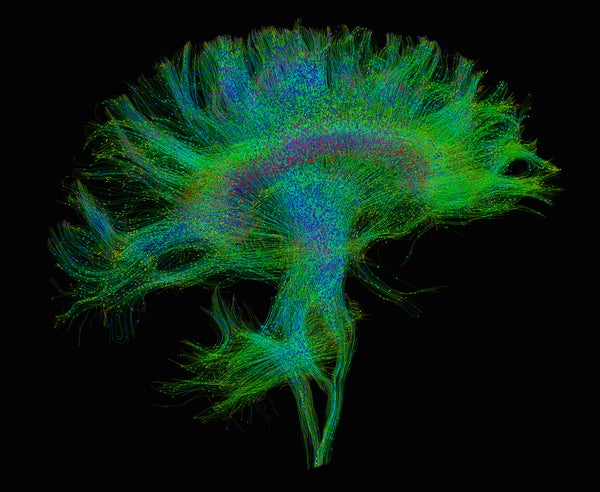
A key tool in these studies is magnetic resonance imaging (MRI)—in particular, a version known as diffusion tensor imaging. This technique can visualize the long fibers that extend out from neurons and link brain regions without having to remove a piece of skull. Like wires, these connections carry electrical information between neurons. And the aggregate of all these links, also known as a connectome, can provide clues about how the brain processes information.
A persistent question about connectomes has to do with what, if anything, distinctive wiring patterns have to do with the evident cognitive differences in a mouse, a monkey or a human. A new methodology called comparative connectomics has identified some general rules of brain wiring across species that may help provide answers. In the meantime, it has also found some unique facets of the human connectome and discovered changes in the cells charged with the upkeep of brain wiring. Together these evolutionary innovations seem to keep information flowing efficiently through a large human brain. And when they are disrupted, they may give rise to psychiatric disorders.
On supporting science journalism
If you're enjoying this article, consider supporting our award-winning journalism by subscribing. By purchasing a subscription you are helping to ensure the future of impactful stories about the discoveries and ideas shaping our world today.
Hypothetically, the most efficient connectome would follow a one-to-many design, with each nerve cell connecting to all of the others. But this approach is prohibitively unworkable because it requires a lot of space to house all these connections and energy to keep them functioning. Alternatively, a one-to-one design, in which each neuron connects to only a single other neuron would be less challenging—but also less efficient: information would have to traverse enormous numbers of nerve cells like stepping-stones to get from point A to point B.
“Real life is in the middle,” says Yaniv Assaf of Tel Aviv University, who published a survey of the connectomes of 123 mammalian species in Nature Neuroscience in June. Assaf came upon his research in a somewhat roundabout way: What began as a weekend hobby of imaging bat brains with his Tel Aviv colleague, Yossi Yovel, turned into a seven-year-long exploration of as many postmortem mammalian brains as they could borrow from a nearby veterinary institute. The investigators looked at a variety of the organs—from the smallest bat brain, which required a magnifying glass to inspect, all the way to the human heavyweight. In between those examples were the brains of giraffes, honey badgers and cows. Among all of them, the team found the same patterns of connections at work: the number of stepping stones to get from one place to another was roughly the same in each of the organs. Differing brains used a similar wiring design.
There were some differences in how this arrangement was achieved, however. Species with few long-range connections linking the two hemispheres of their brain tended to have more short connections within each hemisphere in which nearby areas “talked” intensively with each other. Species with more long-range connections, such as humans and other primates, thinned out these local networks.
This approach to connectivity may reflect geometric constraints on packing a nervous system into a skull. But variations in these links within a species might also track with different abilities. “If you have denser connectivity in one region, you might have a certain ability others wouldn’t,” Assaf says.
Though human brains follow the mammalian connection game plan, they also show some striking innovations. In a head-to-head comparison of human connectomes with those of chimpanzees, our closest living relatives, published last year, Martijn van den Heuvel of Vrije University Amsterdam and anthropologist James Rilling of Emory University revealed 33 human-specific connections. These unique links were longer and more important to network efficiency than 255 connections that were shared in the two species. The distance-spanning connections also tied together high-level “associative” areas in the cortex that are involved in language, tool use and imitation.
“The human brain tends to have a higher investment in keeping those associative areas connected,” van den Heuvel says. This setup could enable efficient integration of information from different parts of the brain, particularly those tasked with conceptual processing. “I think this investment has brought us our more complicated brain functions,” he adds.
When van den Heuvel and his colleagues looked at language areas, a “connectivity fingerprint” popped out. Chimps have their own limited versions of Broca’s and Wernicke’s areas, the regions responsible for human language production and comprehension, respectively. But in humans the connections between the two are stronger. And the connections from Broca’s area to other regions of the brain are actually weaker. It as though the two regions have dedicated their processing might to each other and set the stage for language.
The human-specific connections may form an Achilles’ heel for humans, however. In a study published last November, van den Heuvel, Rilling and their colleagues found human-specific connections were more disrupted in schizophrenia. “This raises the possibility that the evolution of these novel human connections came with a cost,” Rilling says.
While these studies argue for the evolutionary importance of brain connections, the imaging methods are not without mistakes. They have limited resolution, so they may miss a connection ending or taking a turn. This problem means the field needs to draw from other areas of evidence to firm up the findings, says Christine Charvet, an assistant professor at Delaware State University who studies human brain evolution and was not involved in the papers.
Genomics can fill in some of the gaps. A study published in January focused on DNA segments called enhancers, which control whether genes are turned on or off. Menno Creyghton of the Erasmus University Medical Center in the Netherlands and his colleagues found that certain enhancers in humans and chimps differed significantly from those in more distantly related macaques and marmosets. This genomic remodeling took place in cells called oligodendrocytes, which wrap connections with insulating sheaths of protein. Doing so ensures electrical signals quickly reach their destination.
Creyghton suggests the cells are trying to catch up to brain expansion. “These oligodendrocytes need to reinvent themselves to facilitate this larger brain,” he says. “So you have this one spectacular change that gives you a larger brain. And then you need lots of adaptations in the brain to cope with that.”